
Fáìlì:DTI-sagittal-fibers.jpg
Ìtóbi ìkọ́yẹ̀wò yìí: 643 × 600 pixels. Àwọn ìgbéhàn míràn: 257 × 240 pixels | 515 × 480 pixels | 1,021 × 952 pixels.
Fáìlì àtìbẹ̀rẹ̀ (1,021 × 952 pixel, ìtóbi faili: 294 KB, irú MIME: image/jpeg)
|
|
Fáìlì yìí wá láti Wikimedia Commons. Àròyé láti ojúewé ìjúwe rẹ̀ níbẹ̀ nìyí nísàlẹ̀. |
|
Àkótán
| Ìjúwe |
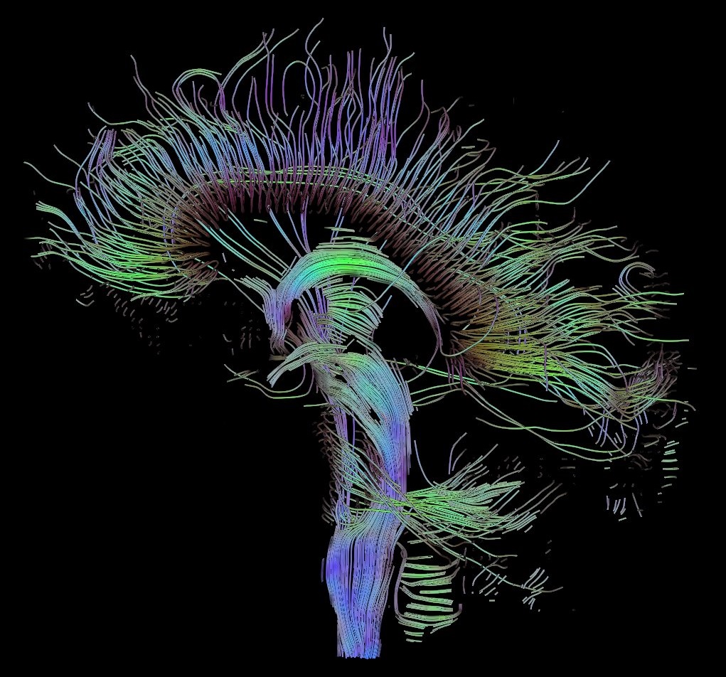
English: Visualization of a DTI measurement of a human brain. Depicted are reconstructed fiber tracts that run through the mid-sagittal plane. Especially prominent are the U-shaped fibers that connect the two hemispheres through the corpus callosum (the fibers come out of the image plane and consequently bend towards the top) and the fiber tracts that descend toward the spine (blue, within the image plane)
Français : Visualisation d'une mesure DTI d'un cerveau humain. Ce qui est représenté sont des faisceaux de fibres reconstruits qui traversent le plan demi-sagittal. On observe les fibres en U qui connectent les deux hémisphères à travers le corps calleux, qui sont particulièrement importantes (les fibres sortent du plan de l'image et par conséquent se courber vers le haut) ainsi que les faisceaux de fibres qui descendent vers la colonne vertébrale (bleu, dans le plan de l'image)
Deutsch: Traktographie-Verfahren rekonstruieren aus den Messdaten der Diffusions-Tensor-Bildgebung den anzunehmenden Verlauf größerer Nervenbahnen. Hier dargestellt sind die Ergebnisse für ein menschliches Gehirn; um die Übersichtlichkeit zu wahren, beschränkt sich die Abbildung auf Bahnen, die die Medianebene schneiden. Insbesondere sind dies die U-förmigen Faserbündel, die die beiden Hirnhälften verbinden (sie durchstoßen die Bildebene und sind nach oben gebogen) sowie die Faserbündel, die zum Rückenmark ziehen (blau dargestellt, liegen innerhalb der Bildebene) |
| Ọjọ́ọdún | |
| Orísun | Iṣẹ́ onítọ̀hún |
| Olùdá | Thomas Schultz |
| Ìyọ̀nda (Ìtúnlò fáìlì yìí) |
Rendering is own work, using a modified version of the BioTensor application developed at the University of Utah. The dataset is courtesy of Gordon Kindlmann at the Scientific Computing and Imaging Institute, University of Utah, and Andrew Alexander, W.M. Keck Laboratory for Functional Brain Imaging and Behaviour, University of Wisconsin, Madison. It is publicly available from [1] |
Ìwé àṣẹ
Èmi gangan, tó jẹ́ pé èmi ni mo ni ẹ̀tọ́àwòkọ iṣẹ́ yìí, fara mọ́ ọ láti tẹ̀ẹ́jáde lábẹ́ àwọn ìwé-àṣẹ ìsàlẹ̀ yìí:

|
Ìyọ̀nda wà láti ṣe àwòkọ, láti pínkàkiri àti/tàbí ṣ'àtúnse ìwé yìí l'ábẹ́ àwọn ọ̀rọ̀ àdéhùn GNU Free Documentation License, Version 1.2 tàbí ìtẹ̀jáde ọjọ́ọwájú lát'ọwọ́ Free Software Foundation; láìsí àwọn Ẹsẹ Aláìyàtọ̀, láìsí àwọn Ọ̀rọ̀-ìwé Níwájú, àti láìsí Ọ̀rọ̀-ìwé Lẹ́yìn. Àwòkọ ìwé àṣẹ náà jẹ́ sísopọ̀ mọ́ abala tí àkọlé rẹ̀ jẹ́ GNU Free Documentation License. |
| Fáìlì yìí wà lábẹ́ ìwé àṣẹ Creative Commons Ìdálórúkọ-Share Alike 3.0 Aláìkówọlé. | ||
| ||
| Àlẹ̀mọ́ ìwé àṣẹ yìí jẹ́ lílẹ̀mọ́ fáìlí yìí gẹ́gẹ́ bíi apá GFDL ìṣọdọ̀tun ìwé àṣẹ. |
Fáìlì yìí wà lábẹ́ ìwé àṣẹ Creative Commons Ìdálórúkọ-Share Alike 2.5 Generic, 2.0 Generic àti 1.0 Generic
- Ẹ ní ààyè:
- láti pín pẹ̀lú ẹlòmíràn – láti ṣàwòkọ, pínkiri àti ṣàgbéká iṣẹ́ náà
- láti túndàpọ̀ – láti mulò mọ́ iṣẹ́ míràn
- Lábẹ́ àwọn àdéhùn wọ̀nyí:
- ìdárúkọ – Ẹ gbọdọ̀ ṣe ọ̀wọ̀ tó yẹ, pèsè ìjápọ̀ sí ìwé-àṣe, kí ẹ sì sọ bóyá ìyípadà wáyé. Ẹ le ṣe èyí lórísi ọ̀nà tó bojúmu, sùgbọ́n tí kò ní dà bii pé oníìwé-àṣe fọwọ́ sí yín tàbí lílò yín.
- share alike – Tó bá ṣe pé ẹ ṣ'àtúndàlú, ṣàyípadà, tàbí ṣ'àgbélé sí iṣẹ́-ọwọ́ náà, ẹ lè ṣe ìgbésíta àfikún yín lábẹ́ ìwé-àṣẹ kannáà tàbí tójọra mọ́ ti àtilẹ̀wa.
Ẹ le ṣàmúyàn ìwé-àṣẹ tí ó wù yín.
Ìtàn fáìlì
Ẹ kan kliki lórí ọjọ́ọdún/àkókò kan láti wo fáìlì ọ̀ún bó ṣe hàn ní àkókò na.
| Ọjọ́ọdún/Àkókò | Àwòrán kékeré | Àwọn ìwọ̀n | Oníṣe | Àríwí | |
|---|---|---|---|---|---|
| lọ́wọ́ | 10:42, 13 Oṣù Kẹ̀wá 2017 | | 1,021 × 952 (294 KB) | Mikael Häggström | Minor crop of black areas at the top and bottom |
| 16:22, 22 Oṣù Kẹ̀sán 2006 |  | 1,021 × 1,125 (203 KB) | Thomas Schultz | {{Information |Description=Visualization of a DTI measurement of a human brain. Depicted are reconstructed fiber tracts that run through the mid-sagittal plane. Especially prominent are the U-shaped fibers that connect the two hemispheres through the corp |
Ìlò fáìlì
Àwọn ojúewé 2 wọ̀nyí únlo fáìlì yí:
Ìlò fáìlì káàkiri
Àwọn wiki míràn wọ̀nyí lo fáìlì yìí:
- Ìlò ní af.wikipedia.org
- Ìlò ní ar.wikipedia.org
- Ìlò ní az.wikiquote.org
- Ìlò ní bn.wikipedia.org
- Ìlò ní cs.wikipedia.org
- Ìlò ní de.wikipedia.org
- Autismus
- Computergrafik
- Bipolare Störung
- Portal:Informatik/Exzellente Artikel
- Portal:Geist und Gehirn/Artikel des Monats
- Diffusions-Tensor-Bildgebung
- Wikipedia:Kandidaten für exzellente Bilder/Archiv2006/17
- Datei:DTI-sagittal-fibers.jpg
- Wikipedia:Exzellente Bilder/Naturwissenschaften
- Portal:Physik/Artikel des Monats 2024-03
- Wikipedia:Exzellente Bilder/Kleine Bilder
- Ìlò ní en.wikipedia.org
- Neurolinguistics
- Tractography
- Portal:Medicine
- User talk:Spikebrennan
- User:Spikebrennan
- Diffusion MRI
- Wikipedia:WikiProject Neuroscience
- Portal:Psychology/Selected article
- Wikipedia:Featured pictures/Sciences/Biology
- Portal:Psychology/Selected article/7
- Wikipedia:Featured pictures thumbs/08
- Wikipedia:Featured picture candidates/DTI-sagittal-fibers.jpg
- Wikipedia:Wikipedia Signpost/2007-11-05/Features and admins
- Wikipedia:Featured picture candidates/November-2007
- Wikipedia:Picture of the day/March 2008
- Connectome
- Template:POTD/2008-03-10
- User talk:Thomas Schultz
- Wikipedia:Wikipedia Signpost/2007-11-05/SPV
- Biological data visualization
- Wikipedia:WikiProject Medicine/Recognized content
- Wikipedia:WikiProject Molecular Biology/Biophysics
- User:Wouterstomp/test
- Wikipedia:WikiProject Anatomy/Resources
- Wikipedia:WikiProject Anatomy/Recognized content
- Wikipedia talk:WikiProject Anatomy/Archive 9
- Portal:Medicine/Recognized content
- User talk:Rhododendrites/Reconsidering FPC on the English Wikipedia
- User:Hydrogenkitsch
- Wikipedia:Wikipedia Signpost/Single/2007-11-05
- Ìlò ní en.wikibooks.org
Ìfihàn ìlò míràn púpọ̀ fún fálì yìí.